
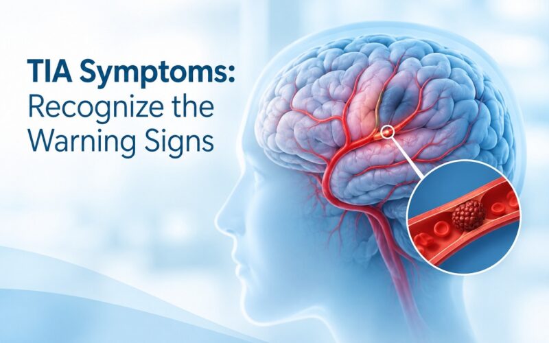
Sudden numbness in an arm, momentary slurring of words, or a fleeting loss of balance can vanish within minutes, tempting you to brush the incident aside. Dismissing these brief episodes is one of the most dangerous medical mistakes a person can make. A temporary disruption in cerebral perfusion is a clear physical indicator that arterial blood flow to your brain has been obstructed.
Understanding a transient ischemic attack is vital because it functions as an early warning for a major ischemic stroke. Recognizing the acute symptoms and receiving immediate diagnostic evaluation saves neural pathways and preserves long-term brain health.
TL;DR: A transient ischemic attack (TIA) is a brief disruption of cerebral blood supply that resolves without leaving permanent tissue damage on standard imaging, but it carries a high risk of an impending stroke. Characterized by sudden facial droop, arm weakness, or speech impairment, a TIA requires emergency hospital evaluation within minutes. High-risk patients are stratified via the ABCD² score and managed with dual antiplatelet therapy, statins, blood pressure control, and surgical revascularization for severe carotid narrowing. Do not wait for symptoms to return; treat every episode as an evolving neurovascular emergency to safeguard long-term brain health.
Quick Answer:- A transient ischemic attack (TIA) is a temporary episode of neurological dysfunction caused by a brief blockage of blood flow to the brain, spinal cord, or retina, occurring without acute infarction (permanent tissue death). While symptoms typically resolve within minutes to hours, a TIA is a critical medical emergency signaling high imminent risk of a debilitating stroke. Immediate emergency hospitalization and neurovascular evaluation are required.
A transient ischemic attack is defined medically as a brief, focal loss of brain, retinal, or spinal function caused by localized vascular ischemia that resolves completely within 24 hours—most often within 15 to 60 minutes—without leaving signs of irreversible parenchymal tissue damage on neuroimaging studies.
What Is a Transient Ischemic Attack and How Does It Occur?
A transient ischemic attack occurs when an arterial branch delivering oxygenated blood and glucose to the brain experiences a temporary occlusion. The primary brain parenchyma demands a relentless vascular supply; it cannot store energy reserves. When a clot restricts capillary perfusion within a specific vascular territory, affected neurons cease normal electrical firing almost immediately.
In a TIA, endogenous fibrinolytic mechanisms break down the microembolus, or collateral blood vessels redirect sufficient flow around the obstruction before cellular energy failure leads to irreversible necrosis. Because perfusion is re-established rapidly, the ischemic penumbra—the rim of threatened tissue surrounding the core—recovers functionality, causing the patient’s noticeable clinical deficits to vanish.
Despite this recovery, a TIA is not a benign occurrence. It proves that the upstream vascular system or the heart is shedding microemboli or that intracranial vessel architecture is compromised by advanced atherosclerotic narrowing.
Atherosclerotic Plaque / Cardiac Thrombus
       ↓
Dislodged Microembolus Travels to Cerebral Artery
       ↓
Transient Microvascular Occlusion & Focal Ischemia
       ↓
Immediate Neurological Deficits (FAST Signs)
       ↓
Endogenous Clot Dissolution / Collateral Reperfusion
       ↓
Resolution of Symptoms Without Structural Infarction
Clinical Summary:-
- Physiological Cause: Transient mechanical obstruction of cerebral or ocular microvasculature.
- Cellular Impact: Reversible cellular ischemia without permanent tissue death (infarction).
- Primary Warning: Indicates an active, unstable vascular or cardiac source capable of triggering a catastrophic stroke.
What Are the Primary Warning Signs and Symptoms of a TIA?
Recognizing the clinical symptoms of a TIA requires vigilance because the manifestations develop abruptly without advance discomfort. The global gold-standard triage tool is the B.E. F.A.S.T. clinical framework:
- B – Balance: Sudden loss of coordination, vertigo, unsteadiness, or ataxia.
- E – Eyes: Sudden monocular blindness (amaurosis fugax), double vision (diplopia), or visual field cuts (hemianopia).
- F – Face: Asymmetrical facial drooping, numbness, or an uneven smile caused by cranial nerve disruption.
- A – Arms: Sudden unilateral muscular weakness, drift, heaviness, or numbness in an arm or leg.
- S – Speech: Dysarthria (slurred speech), expressive aphasia (difficulty producing words), or receptive aphasia (inability to comprehend speech).
- T – Time: Immediate emergency dispatch. The disappearance of symptoms does not cancel the emergency.
Atypical and Gender-Specific Presentations
While motor and speech deficits remain standard across demographics, atypical symptoms can arise, particularly in women, elderly individuals, and diabetic patients. These include:
- Sudden generalized disorientation or acute confusion.
- Sudden sensory hemi-neglect (unawareness of one side of the surrounding environment).
- Transient sensory loss without overt muscular paresis.
- Sudden severe drop attacks without loss of consciousness.
How Does a TIA Differ From an Acute Ischemic Stroke?
While both conditions stem from acute neurovascular interruption, their underlying pathological endpoints, clinical diagnostic markers, and immediate therapeutic goals show key distinctions.
Comparative Analysis: TIA vs. Acute Ischemic Stroke
| Clinical Parameter | Transient Ischemic Attack (TIA) | Acute Ischemic Stroke |
| Tissue Infarction | Absent; no permanent cellular death on MRI-DWI | Present; defined parenchymal necrosis visible on neuroimaging |
| Duration of Symptoms | Typically 5 to 60 minutes; resolves within 24 hours | Persistent; deficits remain long-term or resolve slowly with rehab |
| Emergency Priority | Maximum; aimed at preventing imminent major stroke | Maximum; aimed at immediate salvage of the ischemic penumbra |
| Immediate Therapy | Antiplatelets, anticoagulants, acute blood pressure control | Intravenous thrombolysis (tPA/TNK), mechanical thrombectomy |
| Prognostic Impact | 10–20% risk of full stroke within 90 days if untreated | Substantial risk of long-term cognitive and motor disability |
Transient Ischemia (No Infarction) = TIA
Ischemia + Cell Death (Infarction on DWI) = Stroke
Clinical Summary Box
- Key Difference: Tissue survival vs. permanent parenchymal necrosis.
- Diagnostic Decider: High-resolution Diffusion-Weighted MRI (DWI).
- Triage Rule: Treat every transient episode as an evolving acute stroke until comprehensive emergency imaging proves otherwise.
What Pathological Mechanisms and Risk Factors Trigger a TIA?
A transient ischemic attack is not an isolated disease entity; it is a manifestation of underlying vascular or cardiovascular pathology. The etiological origins generally fall into three broad categories:
        ETIOLOGICAL PATHWAYS OF TIA
 ┌───────────────────┬───────────────────┐
 │                   │                   │
Large-Artery        Cardioembolic       Small-Vessel
Atherosclerosis     Source              Disease
(Carotid/Vertebral) (Afib, PFO, Valve)  (Lacunar Arteriole)
- Large-Artery Atherosclerosis: Progressive accumulation of fibrofatty plaques in the internal carotid or vertebral-basilar arteries. Rupture of a plaque surface exposes collagen, sparking platelet aggregation. These microclots break free and travel downstream to block smaller intracranial arterioles.
- Cardioembolism: Clot formation within cardiac chambers, frequently induced by Atrial Fibrillation (AFib), where erratic atrial contractions permit blood stasis in the left atrial appendage. Other sources include mural thrombi following myocardial infarction, artificial heart valves, or paradoxical emboli moving through a Patent Foramen Ovale (PFO).
- Small-Vessel Occlusion (Lacunar): Longstanding systemic hypertension and chronic hyperglycemia induce lipohyalinosis and microatheroma formation within tiny, deep penetrating cerebral arteries supplying the basal ganglia and internal capsule.
Risk Factor Stratification
- Non-Modifiable: Advanced age (over 55), male biological sex, genetic vascular predisposition, family history of premature stroke.
- Modifiable Medical Risks: Chronic arterial hypertension (the primary driver of vascular remodeling), hyperlipidemia (elevated LDL and ApoB particles), Type 2 Diabetes Mellitus, obstructive sleep apnea, hyperhomocysteinemia.
- Modifiable Behavioral Risks: Tobacco smoking (accelerates endothelial degradation), chronic alcohol abuse, sedentary lifestyle, visceral obesity, high-sodium diets.
How Do Physicians Evaluate and Stratify TIA Severity (The ABCD² Score)?
Emergency physicians and stroke teams use the validated ABCD² clinical prediction score to quantify a patient’s risk of experiencing a completed stroke within 2, 7, and 90 days following an initial TIA.
The ABCD² Clinical Scoring Matrix
| Component | Clinical Finding | Points Assigned |
| A – Age | Age $\ge$ 60 years | 1 point |
| B – Blood Pressure | Systolic $\ge$ 140 mmHg OR Diastolic $\ge$ 90 mmHg at triage | 1 point |
| C – Clinical Features | Unilateral weakness with or without speech impairment | 2 points |
| Speech impairment without unilateral weakness | 1 point | |
| D – Duration | Episode duration $\ge$ 60 minutes | 2 points |
| Episode duration 10 to 59 minutes | 1 point | |
| Episode duration < 10 minutes | 0 points | |
| D – Diabetes | Clinically diagnosed diabetes mellitus | 1 point |
Risk Interpretation and Strategic Action
| Total Score | Risk Category | 2-Day Stroke Risk | Recommended Management |
| 0 – 3 Points | Low Risk | ~1.0% | Rapid outpatient neurovascular clinic workup within 24–48 hours |
| 4 – 5 Points | Moderate Risk | ~4.1% | Hospital admission to a designated acute stroke unit |
| 6 – 7 Points | High Risk | ~8.1% | Immediate critical hospital admission, continuous telemetry, neurovascular imaging |
Clinical Summary Box
- Score Purpose: Quantifies short-term risk of a secondary, completed ischemic stroke.
- Critical Cutoff: A score of 4 or higher demands immediate inpatient hospital admission.
- Limitation: Even low-score patients carry significant vascular risk if underlying high-grade carotid stenosis or atrial fibrillation is discovered.
Which Diagnostic Imaging and Laboratory Workups Are Mandatory?
Determining the source of a transient ischemic attack requires a systematic diagnostic pathway executed within hours of hospital arrival.
                          EMERGENCY TIA WORKUP
                                   │
       ┌───────────────────────────┼───────────────────────────┐
       ▼                           ▼                           ▼
 Neuroimaging                Vascular Assessment         Cardiovascular Evaluation
 (MRI DWI + FLAIR /          (CT Angiography /           (12-Lead ECG / 24-72h Holter /
 Non-Contrast CT)            Carotid Duplex US)          Transthoracic/Transesophageal Echo)
1. Advanced Brain Neuroimaging
- Diffusion-Weighted MRI (DWI): The most sensitive imaging modality. DWI detects microscopic areas of acute cellular edema and restricted water diffusion. If a persistent signal lesion is found, the diagnosis shifts from TIA to a minor acute ischemic stroke.
- Non-Contrast Head CT: Indicated as the initial test if MRI is unavailable, primarily to exclude acute intracranial hemorrhage, subdural hematomas, or intracranial tumors that can mimic TIA symptoms.
2. Neurovascular Vessel Imaging
- CT Angiography (CTA) or Magnetic Resonance Angiography (MRA): Visualizes vascular architecture from the aortic arch through the circle of Willis to detect luminal narrowing, dissection, or intracranial stenoses.
- Carotid Duplex Ultrasound: Assesses hemodynamic flow velocity and measures atherosclerotic plaque burden at the carotid bifurcation.
3. Cardiac and Hematologic Assessments
- 12-Lead Electrocardiogram & Telemetry: Screens for occult arrhythmias, particularly paroxysmal atrial fibrillation. Prolonged 24-hour to 30-day Holter monitoring is used when cardioembolic sources are suspected.
- Echocardiography (TTE/TEE): Transthoracic and transesophageal echocardiograms assess ejection fraction, detect mural thrombi, evaluate valve calcification, and identify patent foramen ovale (PFO).
- Comprehensive Blood Panels: Full blood count, fasting plasma glucose, HbA1c, high-sensitivity lipid profile, renal function, coagulation markers (PT/INR, aPTT), and inflammatory biomarkers (hs-CRP).
What Are the Proven Medical and Surgical Treatments for a TIA?
Treatment for a transient ischemic attack focuses on stabilizing vascular lesions, suppressing platelet aggregation, and managing systemic hemodynamics to prevent secondary ischemic stroke.
       SECONDARY STROKE PREVENTION PROTOCOL
 ┌──────────────────────┬──────────────────────┐
 │                      │                      │
Pharmacological        Interventional         Lifestyle &
Therapy                Procedures             Metabolic Control
(DAPT / DOACs /        (Carotid Endarterectomy/ (BP < 130/80 /
High-Intensity Statins) Stenting)              LDL < 55 mg/dL)
Pharmacological Strategies
1. Dual Antiplatelet Therapy (DAPT)
For patients with non-cardioembolic TIA presenting with high risk (ABCD² $\ge$ 4) or minor stroke, international clinical trials (such as CHANCE and POINT) show that early initiation of Dual Antiplatelet Therapy (Aspirin combined with Clopidogrel) within 24 hours of symptom onset reduces the 90-day risk of recurrent ischemic events compared to aspirin monotherapy.
- Standard Regimen: Clopidogrel loading dose (300–600 mg) plus Aspirin (160–325 mg), followed by Clopidogrel (75 mg/day) and Aspirin (75–81 mg/day) for 21 days, subsequently transitioning to single antiplatelet therapy.
2. Anticoagulation
If cardiac monitoring reveals atrial fibrillation or a ventricular thrombus, antiplatelet agents are replaced with systemic anticoagulation. Direct Oral Anticoagulants (DOACs) such as Apixaban, Rivaroxaban, or Dabigatran are preferred over Vitamin K antagonists (Warfarin) due to their superior safety profiles and lower risk of fatal intracranial hemorrhage.
3. High-Intensity Lipid-Lowering Therapy
Regardless of baseline low-density lipoprotein (LDL) levels, high-intensity statin therapy (Atorvastatin 80 mg or Rosuvastatin 20–40 mg) stabilizes atherosclerotic plaques, reduces vascular inflammation, and improves endothelial reactivity. The target LDL-C is under 55 mg/dL (1.4 mmol/L) for established atherosclerotic disease.
4. Blood Pressure Regulation
In the acute phase, blood pressure is managed cautiously to prevent sudden hypoperfusion of vulnerable penumbral tissue. Long-term goals target resting blood pressure below 130/80 mmHg using ACE inhibitors, ARBs, or thiazide diuretics.
Surgical and Endovascular Interventions
When diagnostic imaging identifies severe symptomatic internal carotid artery stenosis (70% to 99%), mechanical revascularization is indicated to eliminate the embolic source.
Symptomatic Carotid Stenosis
  ├─ 70% – 99% Stenosis ──> Urgent Revascularization (Within 14 days)
  ├─ 50% – 69% Stenosis ──> Individualized Assessment (Age, Sex, Plaque morphology)
  └─ < 50% Stenosis ───────> Aggressive Medical Management
Comparison: Carotid Endarterectomy (CEA) vs. Carotid Artery Stenting (CAS)
| Surgical Feature | Carotid Endarterectomy (CEA) | Carotid Artery Stenting (CAS) |
| Procedural Technique | Open surgical arteriotomy to excise plaque | Minimally invasive endovascular balloon and metallic stent |
| Anesthesia Type | General or regional cervical block | Local anesthesia with conscious sedation |
| Primary Indication | Patients aged $\ge$ 70; standard anatomical risk | Patients with hostile neck anatomy, radiation-induced stenosis, or severe cardiopulmonary comorbidities |
| Associated Risks | Minor cranial nerve palsy, surgical wound hematoma | Slightly higher risk of peri-procedural microembolism |
| Recovery Window | 2 to 4 days hospital stay | 1 to 2 days hospital stay |
Clinical Summary Box
- Primary Medical Step: Rapid deployment of DAPT for 21 days in high-risk non-cardioembolic TIA.
- Primary Surgical Step: Carotid revascularization within 14 days for symptomatic stenosis $\ge$ 70%.
- Metabolic Goal: Achieve blood pressure $< 130/80$ mmHg and LDL $< 55$ mg/dL.
Step-by-Step Emergency Protocol: What Should You Do During an Episode?
When symptoms of a transient ischemic attack appear, every minute matters. Follow this clinical action sequence:
- Do Not Wait for Spontaneous Resolution: Even if the weakness or speech impediment fades after 3 minutes, treat the condition as an evolving stroke.
- Call Emergency Services Immediately: Dial 999 (in the UAE) or your regional emergency dispatch number. Do not attempt to drive yourself or have a family member transport you in a personal vehicle, as ambulances provide trained paramedics and emergency pre-notification to hospital stroke teams.
- Note the Exact Onset Time: Document the precise minute symptoms appeared. This determines candidate eligibility for acute interventions if symptoms escalate into a full-scale stroke.
- Avoid Food, Fluids, and Self-Medication: Never swallow aspirin, antihypertensives, or home remedies before a physician rules out intracranial hemorrhage via neuroimaging. Dysphagia (swallowing impairment) often accompanies acute brain ischemia, creating severe aspiration risks.
- Rest in a Recumbent Position: Keep the patient lying flat with the head slightly elevated (15 to 30 degrees) to optimize cerebral blood flow while guarding against sudden falls or syncopal events.
Real-World Community Insights: Addressing Patient Misconceptions
Insights gathered from patient forums and real-world clinical care reveal recurring misconceptions about transient ischemic attacks.
1. What Most People Misunderstand
- Misconception: “Because my vision and speech returned to normal, the incident was just a bad migraine or exhaustion, so I don’t need a hospital visit.”
- Clinical Reality: Transient resolution indicates that natural clot-dissolving processes barely cleared the vessel in time. The root cause—such as an unstable ulcerated carotid plaque or an atrial thrombus—remains active and untreated, leaving the patient at high risk of a recurrent, disabling stroke within hours or days.
2. Common Mistakes Observed in Patient Recovery
- Discontinuing Antiplatelet Medication: Stopping aspirin or clopidogrel after a few weeks because they “feel fine,” which causes a rebound in platelet reactivity.
- Over-aggressive Blood Pressure Lowering at Home: Taking extra doses of antihypertensives during an attack, which lowers cerebral perfusion pressure and worsens brain ischemia.
- Failing to Track Occult Atrial Fibrillation: Skipping follow-up cardiac monitoring after normal hospital ECGs miss paroxysmal arrhythmias.
3. What Experienced Patients and Neurologists Recommend
- Keep an Emergency Medical Card: Carry a printed list of current medications, baseline blood pressure measurements, and your primary stroke center’s emergency phone number.
- Adopt Home Blood Pressure Logging: Record seated morning and evening blood pressure measurements to ensure therapies maintain readings below target thresholds.
- Undergo Comprehensive Lifestyle Modification: Pair prescribed pharmacotherapy with regular cardiovascular exercise, smoking cessation, and Mediterranean-style nutrition.
People Also Ask
Can an MRI show a TIA?
By traditional definition, a true transient ischemic attack does not leave permanent tissue damage, meaning standard CT scans and conventional MRI sequences will show normal brain parenchyma. However, modern Diffusion-Weighted Magnetic Resonance Imaging (DWI) detects subtle cellular injury in approximately 30% to 50% of patients diagnosed clinically with a TIA. When DWI shows an acute focal lesion, the event is reclassified as a minor ischemic stroke with transient symptoms.
How long does a transient ischemic attack usually last?
Most transient ischemic attacks last between 2 and 15 minutes, with the average duration sitting around 10 minutes. While the traditional diagnostic definition allows up to 24 hours for symptom resolution, episodes persisting past one hour typically cause microscopic tissue necrosis, reflecting an evolving acute ischemic stroke rather than an uncomplicated TIA.
Can high blood pressure cause a mini-stroke?
Yes, chronic high blood pressure is the single most significant modifiable cause of a mini-stroke. Sustained arterial hypertension causes vascular endothelial injury, accelerates carotid atherosclerosis, and damages small penetrating cerebral blood vessels through lipohyalinosis. Sudden blood pressure spikes can also destabilize arterial plaques, causing microemboli to travel downstream and block cerebral arteries.
What is the life expectancy after a transient ischemic attack?
Life expectancy following a transient ischemic attack depends on prompt medical intervention and vascular risk management. Studies indicate that without medical treatment, roughly 10% to 20% of TIA patients suffer a major stroke within 90 days, and long-term cardiovascular mortality increases. However, with modern secondary prevention—including antiplatelets, statins, blood pressure control, and surgical carotid revascularization when appropriate—patients can achieve long-term survival rates comparable to those of individuals without cerebrovascular disease.
Is a TIA painful?
No, a transient ischemic attack is rarely painful. Brain tissue contains no sensory pain receptors, meaning ischemia itself causes no headache or physical ache. Symptoms typically manifest as painless functional losses, such as sudden weakness, numbness, speech difficulties, or visual field deficits. Mild headaches occur in a small minority of cases, but severe head pain is more characteristic of hemorrhagic strokes, arterial dissections, or migraine auras.
Frequently Asked Questions (FAQs)
How can I tell if my symptoms are from a TIA or a complex migraine aura?
A migraine aura typically develops gradually over 5 to 20 minutes, producing “positive” visual disturbances such as shimmering zig-zag lines, flashing spots (scintillating scotoma), or tingling sensations that slowly spread across limbs before a headache begins. In contrast, a transient ischemic attack develops instantaneously within seconds, causing “negative” neurological deficits such as loss of vision, loss of sensation, or sudden muscular paralysis. A TIA rarely features spreading visual phenomena or severe subsequent headaches. Because clinical features can overlap in older adults, urgent neurovascular imaging and a specialist medical evaluation are necessary to rule out cerebral ischemia.
What lifestyle modifications help prevent another TIA?
Preventing recurrent vascular attacks requires aggressive, sustainable lifestyle changes alongside medical therapy. Patients should adopt a Mediterranean or DASH diet high in leafy greens, extra-virgin olive oil, whole grains, and lean proteins while eliminating processed foods and restricting daily sodium intake to under 1,500 mg. Ceasing tobacco use is mandatory, as smoking immediately damages endothelial linings and increases platelet aggregability. Engage in at least 150 minutes of moderate aerobic exercise weekly once cleared by your neurologist, minimize alcohol intake, and maintain healthy sleep hygiene to prevent worsening sleep apnea.
Can stress directly trigger a transient ischemic attack?
While psychological stress itself rarely causes a TIA directly, acute emotional stress causes a sharp rise in sympathetic nervous system activity. This surges adrenaline, increases heart rate, and causes acute spikes in systemic blood pressure. In patients with underlying atherosclerosis or unstable plaques, this sudden hemodynamic stress can rupture a plaque or trigger paroxysmal atrial fibrillation, causing microclots to form and travel to the brain. Chronic stress also promotes systemic inflammation, which accelerates long-term cerebrovascular disease.
Why is dual antiplatelet therapy prescribed immediately after a high-risk TIA?
Dual Antiplatelet Therapy (DAPT)—most commonly combining Aspirin with Clopidogrel—is prescribed immediately following a high-risk TIA (ABCD² score $\ge$ 4) because clinical trials prove it reduces the risk of a recurrent, disabling ischemic stroke during the critical 21-day high-risk period. Aspirin inhibits the cyclooxygenase-1 (COX-1) pathway, while Clopidogrel blocks the P2Y12 adenosine diphosphate (ADP) receptor on platelets. Inhibiting these two complementary pathways provides greater protection against platelet aggregation and thrombus propagation than either medication alone. After 21 days, DAPT is typically tapered down to single antiplatelet monotherapy to minimize long-term bleeding risks while maintaining vascular protection.
What is amaurosis fugax, and why is it linked to a TIA?
Amaurosis fugax is a temporary, painless loss of vision in one eye, often described as a dark shade or curtain dropping vertically over the visual field. It occurs when a microembolus—typically shed from an atherosclerotic plaque located at the carotid artery bifurcation—temporarily occludes the central retinal artery or ophthalmic artery. Because the ophthalmic artery is the first major branch of the internal carotid artery, amaurosis fugax serves as a clear warning of significant ipsilateral carotid stenosis, requiring urgent duplex ultrasonography and neurovascular imaging.
What driving restrictions apply after experiencing a TIA?
Driving regulations post-TIA vary by jurisdiction and clinical recovery. In most regions, patients are legally restricted from driving for at least two to four weeks following symptom resolution. This waiting period allows the medical team to complete comprehensive diagnostic workups, begin secondary prevention therapies, and verify that no recurrent neurological deficits or underlying cardiac arrhythmias go unmanaged. Commercial drivers face stricter regulations and require detailed clearance from a certified neurologist. Always consult your attending physician regarding local driving laws.
Can a transient ischemic attack cause permanent cognitive changes or memory loss?
Although a single TIA does not leave a large area of permanent tissue damage on conventional imaging, recurrent TIAs or chronic cerebral microvascular disease can lead to vascular cognitive impairment over time. Patients often report transient fatigue, mental fogginess, or heightened anxiety in the weeks following an attack. Furthermore, if modern diffusion-weighted MRI detects micro-infarctions, subtle cognitive changes may persist. Maintaining blood pressure control, lowering cholesterol, and treating obstructive sleep apnea are vital to preventing cumulative cerebral damage and long-term vascular dementia.
Experiencing a transient ischemic attack is a pivotal medical warning that should never be ignored. The temporary disappearance of neurological deficits does not mean the underlying vascular threat has passed; it simply means you have a critical window to prevent a life-altering ischemic stroke. Modern stroke centers combine rapid diagnostic imaging, cardiac monitoring, and preventive therapies to locate the clot source and restore vascular health. By taking immediate emergency action, undergoing a thorough medical workup, and following targeted medical advice, you can protect your cognitive independence and lower your long-term cardiovascular risk.
Key Takeaways
- Recognize the FAST Signs: Drooping face, arm weakness, and slurred speech demand an immediate 999 emergency call.
- Treat Every Incident as an Emergency: Resolving symptoms do not mean the danger is gone; the clot source remains active.
- Understand the 21-Day Window: High-risk TIAs require immediate dual antiplatelet therapy (Aspirin plus Clopidogrel) to stop secondary strokes.
- Examine the Carotid Arteries: Significant stenosis ($\ge$ 70%) requires urgent evaluation for surgical or endovascular revascularization.
- Track Atrial Fibrillation: Occult cardiac arrhythmias must be ruled out with prolonged Holter monitoring.
Actionable Next Steps
- Call Emergency Services Immediately: Dial 999 in the UAE if you or someone nearby experiences sudden neurological deficits.
- Undergo Brain and Vascular Imaging: Ensure an emergency department performs an MRI with Diffusion-Weighted Imaging (DWI) and vascular imaging (CTA/MRA).
- Obtain an ABCD² Score Evaluation: Work with a neurologist to quantify your 48-hour stroke risk and determine if hospital admission is necessary.
- Adhere Strictly to Medication: Take all prescribed antiplatelet or anticoagulant medications consistently, never discontinuing them without specialist supervision.
- Schedule a Comprehensive Follow-up: Book a consultation with a stroke neurologist, complete prolonged Holter monitoring, and implement a personalized cardiovascular lifestyle plan.
رعاية السكتة الدماغية في كليفلاند كلينك أبوظبي (Abu Dhabi Healthcare Services)
يُعتبر معهد الأعصاب التخصصي ومركز السكتة الدماغية المعتمد في مستشفى كليفلاند كلينك أبوظبي (Cleveland Clinic Abu Dhabi) الوجهة الرائدة في دولة الإمارات العربية المتحدة لتقديم الرعاية الطارئة والتشخيص المتقدم لحالات النوبة الإقفارية العابرة والسكتات الدماغية؛ حيث يضم المستشفى أحدث تقنيات التصوير بالرنين المغناطيسي وتصوير الأوعية الدموية العصبية، إلى جانب نخبة من استشاريي الأعصاب وجراحة الأوعية الدقيقة لضمان الوقاية السريعة من السكتات الدماغية الدائمة. يقع المستشفى في موقع استراتيجي في 59 شارع حموده بن علي الظاهري، جزيرة الماريه، أبوظبي، الإمارات العربية المتحدة، ويقدم قسم الطوارئ خدماته على مدار 24 ساعة يومياً طوال أيام الأسبوع لإنقاذ حياة المرضى. للحصول على الدعم وحجز المواعيد مع كبار الأطباء، يمكنكم زيارة الموقع الإلكتروني الرسمي https://www.clevelandclinicabudhabi.ae أو الاتصال مباشرة على الرقم المجاني 80082223 (أو 97180082223+)، كما يمكنكم الوصول المباشر والاطلاع على التقييمات عبر البحث عن ملفنا على خرائط جوجل Google Business Profile للوصول السريع إلى المستشفى في الحالات العصبية والحرجة.